As part of a bryology course at the University of British Columbia, I looked at some of the sphagnum mosses in Camosun Bog. I'd like to show you one of the mosses,
Sphagnum palustre, magnified up close. This is what is looks like fresh.
If I were to pull out one individual and dry it, this is what it would look like:
Its hard to see, but there are two kind of "leaves" on this moss, ones that go down the stem and ones that come off on horizontal branches.
From Crum, Howard. 1984. North American Flora. Series II. Part 11. Sphagnopsida. Sphagnaceae. Published by the New York Botanical Garden, Bronx, NY.
This is what a stem leaf looks like and what the stem leaf cells look like:
There are two types of cells. One type is green and undergoes photosynthesis. The other type is clear and allows the plant to take up water as a storage reservoir.
This is what a divergent leaf looks like under the microscope. In this case, there are holes where water can enter the cell.
Now if we did a cross-section of the leaf, what would it look like?
The leaf is only one cell thick! Here you can really see how photosynthetic cells are squeezed between transparent cells. Next is a cross-section of the stem.
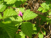
The big clear cells are designed to store water. The closely packed cells in the middle are designed to move water from the bottom of the plant, to the top. There are no photosynthetic cells. Some sphagnums in the bog can be identified by the eye, like the bright red
S. capillifolium, while others require comparing structures under the microscope, like I've just shown. Sphagnum is very pretty both to the naked eye, as well as under a microscope.